How to perform a Gram stain


  One of the most important techniques in bacteriology is the Gram stain, first described by Hans-Christian Gram in 1884. Scientists still do not understand fully how this stain works but it allows the differentiation of bacteria into two groups: Gram-positive bacteria can retain a complex made between crystal-violet and iodine when decolourised with acetone, whereas Gram-negative bacteria are decolourised on treatment with acetone.

This property reveals fundamental differences in the cell envelope between the two groups. Gram-positive bacteria have many layers of peptidoglycan in their cell wall; Gram-negative bacteria have only one or two peptidoglycan layers but, additionally, they have an outer membrane. These differences have important consequences. For example, certain antibiotics cannot penetrate the outer membrane of Gram-negative bacteria, which are intrinsically resistant to these drugs as a consequence.

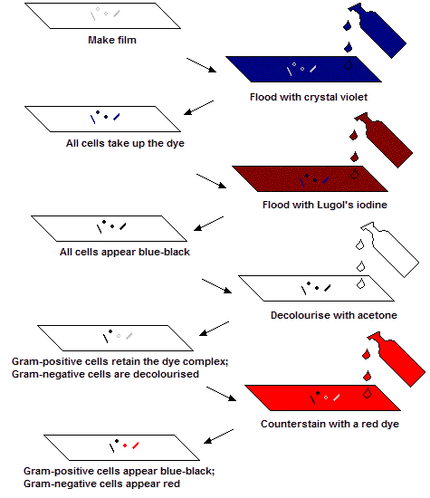
Below is a schematic, showing how the Gram stain works:

Steps in the Gram staining reaction
  As well as differentiating cells on the basis of the Gram reaction, the Gram stain enables the shape of bacterial cells to be observed easily. Bacteria fall into two major classes: rod shaped bacteria are known as 'bacilli' (singular: 'bacillus') and round or roundish bacteria are known as 'cocci' (singular: 'coccus').

If you have trouble remembering which is which, just think of 'bacillus' as a rod-shaped word and 'coccus' as a round word...